
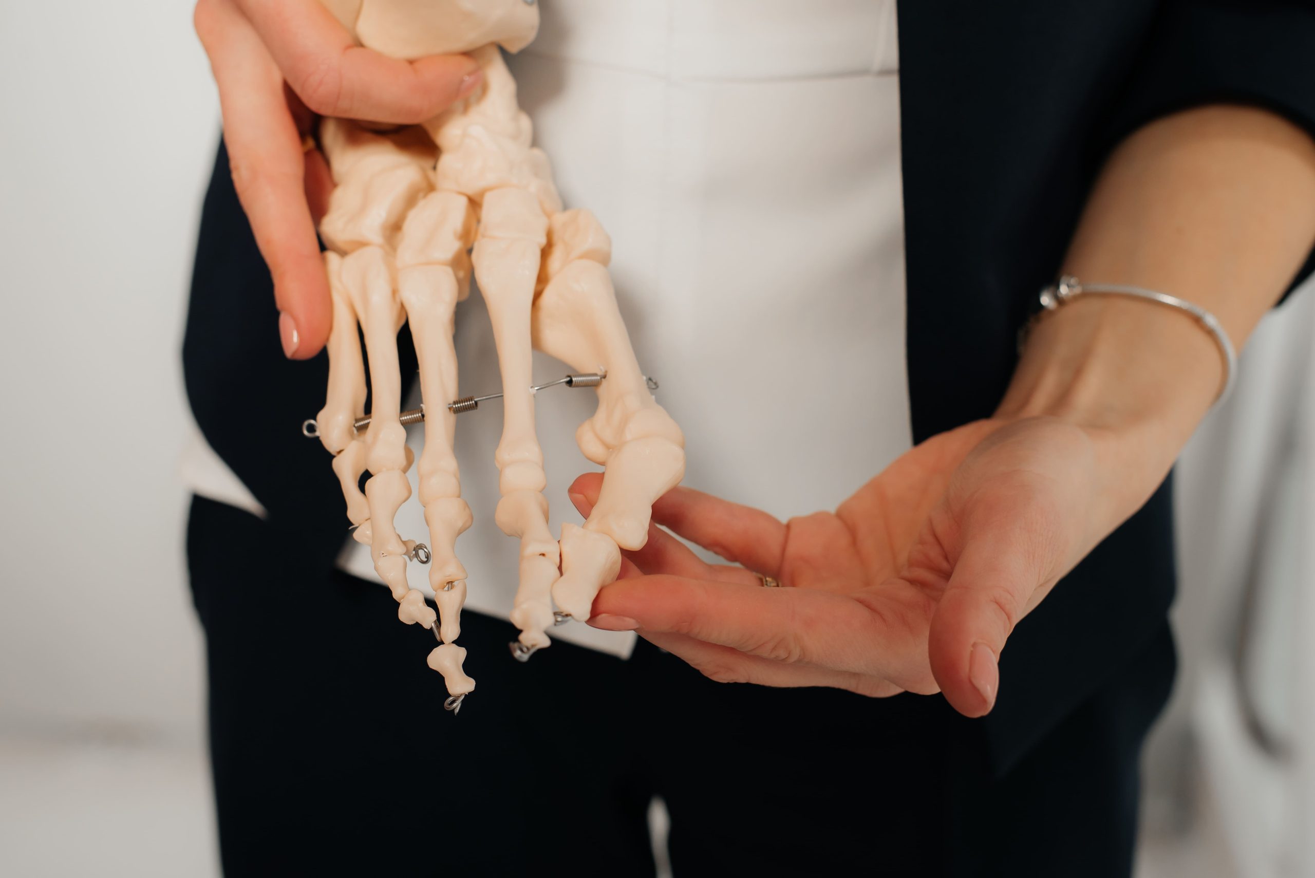
Một vết gãy trông giống như bong gân nhẹ trên phim X-quang ban đầu có thể âm thầm phá hủy cổ tay trong nhiều tháng hoặc nhiều năm. Nằm ở gốc ngón cái, xương thuyền nhỏ, hình hạt điều thường thoát khỏi việc phát hiện sớm, khiến nó trở thành một trong những chấn thương cổ tay thường bị bỏ sót nhất.
Điều khiến vết gãy “ẩn” này trở nên đặc biệt nguy hiểm là nguồn cung cấp máu ngược dòng độc đáo của nó, nghĩa là một vết gãy có thể dễ dàng cắt đứt tuần hoàn quan trọng và gây chết xương lâu dài nếu không có sự can thiệp sớm của bác sĩ chuyên khoa tay.
Cách Xương Thuyền Bị Gãy
Xương thuyền rất dễ bị gãy trong các tác động đột ngột do vị trí của nó là cầu nối chính giữa bàn tay và cẳng tay.
- Ngã Chống Tay (FOOSH): Nguyên nhân phổ biến nhất là ngã chống tay với cổ tay bị duỗi quá mức, dồn toàn bộ trọng lượng cơ thể trực tiếp qua lòng bàn tay.
- “Eo” Xương Dễ Tổn Thương: Khi chuẩn bị ngã, phản xạ bảo vệ bản năng mở lòng bàn tay, tập trung ứng suất cơ học tối đa vào phần hẹp nhất, dễ vỡ nhất ở giữa xương.
- Nguy Cơ Tác Động Mạnh: Tai nạn trong các môn thể thao va chạm, tai nạn xe đạp và ngã tại nơi làm việc là những nguyên nhân hàng đầu gây ra chấn thương đặc thù này.
- Yếu Tố Tuổi Tác: Người trẻ tuổi bị gãy xương thuyền thường xuyên hơn vì xương đặc của họ truyền lực hiệu quả, trong khi người lớn tuổi có mật độ xương thấp hơn thường dễ bị gãy đầu xa của xương quay, một trong những xương cẳng tay tạo thành khớp cổ tay, hơn là gãy xương thuyền.
Nhận Biết Triệu Chứng Gãy Xương Thuyền
Bởi vì gãy xương thuyền thường trông và cảm giác giống như bong gân cổ tay thông thường, việc xác định các triệu chứng khu trú tinh tế là rất quan trọng để ngăn ngừa tổn thương khớp lâu dài. Cơn đau thường tăng lên khi thực hiện động tác nắm hoặc xoay ngón cái, kèm theo sưng nhẹ chỉ giới hạn ở gốc ngón cái chứ không phải sưng lan rộng.
Kiểm Tra Hố Giải Phẫu và Hơn Thế
Đánh giá lâm sàng dựa trên các xét nghiệm chèn ép khu trú cụ thể, chẳng hạn như nén trục ngón cái hoặc ấn vào mấu xương nổi ở gốc lòng bàn tay, để xác định chính xác chấn thương xương thuyền. Khi các thao tác nhắm mục tiêu này gây ra đau, điều đó cho thấy mạnh mẽ một vết gãy và giải thích tại sao bệnh nhân bị yếu đột ngột khi thực hiện các công việc hàng ngày cơ bản như vặn chìa khóa hoặc xách túi.
Tại Sao X-quang Tiêu Chuẩn Bỏ Sót Gãy Xương Thuyền
X-quang tiêu chuẩn ban đầu thường bỏ sót gãy xương thuyền vì hình dạng không đều, giống hạt điều của xương có thể che giấu các vết nứt tóc. Ngay cả khi bác sĩ chuyên khoa yêu cầu chụp X-quang ‘góc nhìn xương thuyền’ chuyên dụng—nơi cổ tay được đặt ở một góc cụ thể—đường gãy có thể vẫn vô hình trong 10 đến 14 ngày đầu cho đến khi xương bắt đầu tiêu hủy xung quanh vết gãy. Khi nghi ngờ có vết gãy nhưng X-quang rõ ràng, các phương pháp chụp tiên tiến như MRI hoặc CT là cần thiết để phát hiện tổn thương ẩn và hướng dẫn lập kế hoạch phẫu thuật.
Vấn Đề Cung Cấp Máu
Bởi vì máu vào xương thuyền chủ yếu qua các mạch dọc theo gờ lưng của nó gần eo và di chuyển theo hướng ngược dòng từ xa đến gần, một vết gãy qua eo có thể cắt đứt tuần hoàn đến cực gần. Bị thiếu hụt các chất dinh dưỡng quan trọng, mảnh xương bị cô lập này có thể bị hoại tử vô mạch (chết xương), khiến nó xẹp xuống dưới áp lực cổ tay bình thường và gây ra viêm khớp đau đớn vĩnh viễn.
Nguy cơ hoại tử vô mạch liên quan trực tiếp đến vị trí gãy xương. Cực gần mang nguy cơ cao nhất, với tỷ lệ lên đến 100% được báo cáo do sự phụ thuộc của nó vào dòng máu trong xương ngược dòng — một nguy cố hữu với vị trí này và không chỉ phụ thuộc vào việc vết gãy có được điều trị hay không. Gãy một phần ba giữa (eo) mang nguy cơ trung bình khoảng 30%, trong khi gãy một phần ba xa hiếm khi dẫn đến hoại tử vô mạch.
Không Liền Xương và Hậu Quả Của Nó
Khi gãy xương thuyền không lành do lưu lượng máu kém, di lệch vết gãy hoặc điều trị chậm trễ, nó dẫn đến một tình trạng gọi là không liền xương. Nguy cơ không liền xương là đáng kể với các vết gãy di lệch, đạt từ 14% đến 50% hoặc cao hơn nếu vết gãy không được điều trị hoặc quản lý không đầy đủ. Nếu không được điều trị, vết gãy không lành này kích hoạt một mô hình suy thoái cổ tay có thể dự đoán được gọi là SNAC (xẹp tiến triển do không liền xương thuyền), phá hủy vĩnh viễn sụn và hạn chế chuyển động cổ tay.
Phác Đồ Điều Trị Cho Gãy Xương Thuyền
Tùy thuộc vào vị trí cụ thể và mức độ nghiêm trọng của vết gãy, các bác sĩ chuyên khoa chỉnh hình sẽ khuyến nghị bó bột bảo vệ nghiêm ngặt hoặc phẫu thuật cố định để đảm bảo xương lành đúng cách.
Cố Định Bằng Bột
Đối với các vết gãy sạch, không di lệch, một loại bột spica ngón cái chuyên dụng được áp dụng để giữ cổ tay hoàn toàn bất động và bảo vệ xương đang lành dễ tổn thương. Thời gian bó bột thay đổi đáng kể tùy thuộc vào vị trí gãy: gãy cực xa có thể lành trong khoảng sáu tuần, gãy eo thường cần tám đến mười hai tuần, và gãy cực gần có thể cần mười hai tuần hoặc lâu hơn, được hướng dẫn bằng chụp ảnh liên tiếp để xác nhận liền xương trước khi tháo bột.
Cố Định Bằng Phẫu Thuật
Khi các vết gãy bị di lệch hoặc nằm ở cực gần nguy cơ cao, bác sĩ phẫu thuật bàn tay thường sử dụng một vít nén nhỏ, không đầu để cố định cơ học các mảnh xương. Phương pháp cố định bên trong này giúp tránh nhu cầu bó bột lâu dài, mang lại sự trở lại các hoạt động hàng ngày nhanh hơn và dễ dự đoán hơn cho các vận động viên và chuyên gia làm việc.
⚠️ Lưu Ý Quan Trọng: Tiếp tục sử dụng một cổ tay có khả năng bị gãy làm chậm chẩn đoán và tăng nguy cơ biến chứng. Nếu bạn bị đau cổ tay dai dẳng sau khi ngã, ngay cả với X-quang ban đầu bình thường, hãy yêu cầu chụp ảnh theo dõi hoặc đánh giá bác sĩ chuyên khoa trong vòng hai tuần.
Phục Hồi và Tái Huấn Luyện
Đạt được sự liền xương thích hợp chỉ là cột mốc đầu tiên, vì nhiều tháng bất động thường khiến bệnh nhân phải vật lộn với cứng khớp cổ tay và mất sức nắm đáng chú ý. Trị liệu bàn tay chuyên dụng là rất quan trọng trong giai đoạn này để chuyển đổi an toàn khớp từ các chuyển động biên độ nhẹ nhàng sang các bài tập kháng lực có mục tiêu. Trong khi các vết gãy được cố định bằng phẫu thuật thường cho phép trở lại các công việc nhẹ trong vòng sáu tuần, các chấn thương điều trị bằng bột thường đòi hỏi một giai đoạn phục hồi chức năng dài hơn, chuyên sâu hơn do teo cơ kéo dài.
Trở Lại Hoạt Động
Việc tiếp tục thể thao hoặc lao động chân tay nặng nhọc đòi hỏi một cách tiếp cận thận trọng, tăng dần, thường sử dụng nẹp bảo vệ tùy chỉnh để bảo vệ xương đang lành khỏi tái chấn thương đột ngột. Các vết gãy không biến chứng thường thấy sức nắm phục hồi đến mức gần như bình thường trong vòng sáu tháng, mặc dù các trường hợp phức tạp liên quan đến chết xương trước đó hoặc các mảnh không liền sẽ cần một thời gian phục hồi dài hơn nhiều.
Ngăn Ngừa Chẩn Đoán Chậm Trễ
Mẹo Nhanh
Sau bất kỳ cú ngã nào chống tay, hãy tự kiểm tra hố giải phẫu của bạn bằng cách ấn mạnh vào chỗ lõm ở gốc ngón cái trong khi gập cổ tay. Đau đáng kể cần được đánh giá y tế ngay cả khi X-quang ban đầu có vẻ bình thường.
Khi Nào Cần Tìm Kiếm Sự Giúp Đỡ Chuyên Nghiệp
- Đau cổ tay dai dẳng kéo dài hơn vài ngày sau khi ngã
- Đau ở chỗ lõm tại gốc ngón cái
- Đau khi nắm đồ vật hoặc xoay cổ tay
- Sưng hoặc bầm tím ở gốc ngón cái
- Không có khả năng chịu trọng lượng hoặc đẩy qua bàn tay bị thương
Recognising Scaphoid Fracture Symptoms
Because scaphoid fractures often look and feel like a standard wrist sprain, identifying the subtle, localised symptoms is critical to preventing long-term joint damage. Pain typically intensifies during gripping movements or thumb rotation, accompanied by minor puffiness strictly confined to the base of the thumb rather than widespread swelling.
The Snuffbox Test and Beyond
Clinical evaluations rely on specific localised pressure tests, such as compressing the thumb’s axis or pressing the bony prominence at the base of the palm, to accurately pinpoint a scaphoid injury. When these targeted manoeuvres trigger pain, it strongly suggests a fracture and explains why patients experience sudden weakness when performing basic daily tasks like turning keys or holding bags.
Why Standard X-Rays Miss Scaphoid Fractures
Initial standard X-rays frequently miss scaphoid fractures because the bone’s irregular, cashew-like shape can hide hairline cracks. Even when a specialist orders dedicated ‘scaphoid view’ X-rays—where the wrist is positioned at a specific angle—the fracture line may remain invisible for the first 10 to 14 days until the bone begins to resorb around the break. When a break is suspected but X-rays are clear, advanced scans like an MRI or CT are essential to uncover the hidden damage and guide surgical planning.
The Blood Supply Problem
Because blood enters the scaphoid primarily through vessels along its dorsal ridge near the waist and travels in a retrograde direction from distal to proximal, a fracture through the waist can cut off circulation to the proximal pole. Starved of vital nutrients, this isolated bone fragment can suffer avascular necrosis (bone death), causing it to collapse under normal wrist pressure and trigger permanent, painful arthritis.
The risk of avascular necrosis is directly linked to fracture location. The proximal pole carries the highest risk, with rates of up to 100% reported due to its reliance on retrograde intraosseous blood flow — a risk that is inherent to this location and not solely dependent on whether the fracture is treated. Middle-third (waist) fractures carry a moderate risk of approximately 30%, while distal third fractures rarely result in avascular necrosis.
Non-Union and Its Consequences
When a scaphoid fracture fails to heal due to poor blood flow, fracture displacement, or delayed treatment, it results in a condition known as non-union. The risk of non-union is substantial with displaced fractures, reaching 14% to 50% or higher if the fracture is untreated or inadequately managed. Left untreated, this non-healing break triggers a predictable pattern of wrist deterioration called SNAC (scaphoid non-union advanced collapse), which permanently destroys cartilage and restricts wrist movement.
Treatment Pathways for Scaphoid Fractures
Depending on the specific location and severity of the break, orthopaedic specialists will recommend either strict cast protection or surgical stabilisation to ensure the bone heals properly.
Cast Immobilisation
For clean, non-displaced fractures, a specialised thumb spica cast is applied to keep the wrist completely still and protect the delicate healing bone. Casting duration varies considerably depending on fracture location: distal pole fractures may heal in approximately six weeks, waist fractures typically require eight to twelve weeks, and proximal pole fractures may require twelve weeks or longer, guided by serial imaging to confirm union before cast removal.
Surgical Fixation
When fractures are displaced or located in the high-risk proximal pole, a hand surgeon will typically use a tiny, headless compression screw to mechanically stabilise the bone fragments. This internal fixation bypasses the need for long-term casting, offering a faster and more predictable return to daily activities for athletes and working professionals.
⚠️ Important Note: Continuing to use a potentially fractured wrist delays diagnosis and increases complication risk. If you experience persistent wrist pain after a fall, even with normal initial X-rays, request follow-up imaging or specialist evaluation within two weeks.
Recovery and Rehabilitation
Achieving proper bone union is simply the first milestone, as months of immobility often leave patients struggling with wrist stiffness and a noticeable loss of grip strength. Dedicated hand therapy is crucial during this phase to safely transition the joint from gentle range-of-motion movements to targeted resistance exercises. While surgically stabilised fractures often allow a return to light tasks within six weeks, cast-treated injuries typically demand a longer, more intensive rehabilitation period due to prolonged muscle wasting.
Returning to Activities
Resuming sports or demanding physical labour requires a cautious, graduated approach, often utilising custom protective splints to safeguard the healing bone from sudden re-injury. Uncomplicated fractures generally see grip strength recover to near-normal levels within six months, though complex cases involving prior bone death or non-healing fragments will require a much longer recovery timeline.
Preventing Delayed Diagnosis
Quick Tip
After any fall onto an outstretched hand, test your own anatomical snuffbox by pressing firmly into the hollow at the base of your thumb while bending your wrist. Significant tenderness warrants medical evaluation even if initial X-rays appear normal.
When to Seek Professional Help
- Persistent wrist pain lasting more than several days after a fall
- Tenderness in the hollow at the base of your thumb
- Pain when gripping objects or turning your wrist
- Swelling around the thumb side of your wrist
- Weakness when pinching or grasping
- Wrist pain that improves but returns with activity
- Previous wrist injury that never fully resolved
Commonly Asked Questions
Can a scaphoid fracture heal on its own without treatment?
Untreated scaphoid fractures occasionally heal spontaneously, but the risk of non-union and subsequent complications makes this approach inadvisable. The scaphoid’s limited blood supply means fractures generally require either strict immobilisation or surgical stabilisation to achieve reliable healing. Delaying treatment increases the likelihood of non-union, which requires more complex surgery to address.
How do I know if my wrist sprain might actually be a scaphoid fracture?
Scaphoid fracture symptoms overlap substantially with wrist sprains, making clinical distinction difficult. Key differences include tenderness specifically in the anatomical snuffbox, pain with thumb movements, and symptoms persisting beyond a week. Sprains typically improve steadily, while fractures cause pain that plateaus or worsens with activity.
Why might my doctor recommend surgery for a fracture that looks minor on X-ray?
Fracture location influences treatment decisions more than apparent severity. Proximal pole fractures carry high non-union and avascular necrosis risk regardless of displacement, making surgery a more reliable option in some cases. Additionally, patient factors such as occupation, activity level, and inability to tolerate prolonged casting may favour surgical treatment for fractures that would otherwise qualify for conservative management.
What happens if a scaphoid fracture is not diagnosed for several months?
Delayed diagnosis allows the fracture to progress towards non-union. The bone ends may develop fibrous tissue or cartilage instead of bridging with new bone. Treatment at this stage requires surgery to remove the abnormal tissue, apply a bone graft to stimulate healing, and stabilise the fragments with internal fixation. Outcomes may remain favourable but require more extensive intervention.
How long before I can return to sports after a scaphoid fracture?
Return-to-sport timelines vary by treatment method and sport demands. Surgically treated fractures in non-contact athletes may allow return in three months. Contact sports and activities with fall risk typically require four to six months, regardless of treatment method. Your surgeon will use imaging and clinical examination to determine readiness rather than relying solely on time elapsed.
Next Steps
Scaphoid fractures diagnosed within the first week heal more reliably than those identified after delays of several weeks or months. If initial X-rays are negative but snuffbox tenderness, reduced grip strength, or pain with thumb movement persists, follow-up imaging, such as MRI or CT, is required to rule out a fracture. Fracture location, particularly proximal pole involvement, determines whether casting or surgical fixation is the appropriate course.
If you are experiencing tenderness in the anatomical snuffbox, persistent pain when gripping objects, or wrist symptoms that have not resolved after a fall, a hand surgeon can assess for scaphoid fracture and arrange appropriate imaging to guide treatment.

Don't Let Hand Pain Affect Your Life
Restore Function and Improve Your Quality of Life
Our clinic provides a comprehensive, one-stop service for diagnosis and treatment.
Make An Enquiry